Optical Coherence Tomography (OCT) is a non-invasive optical imaging modality that provides high-resolution real-time 3D images. Using an interferometry technique, OCT can differentiate the back-scattered light from different layers within the sample and reconstruct the microstructure of tissue. The resolution of an OCT system depends on the coherence length of the light source. With a low-coherence light source (i.e. shorter center wavelength and broader bandwidth), cellular images with an axial resolution lower than one micron can be achieved. (Fig. 1)
Optical Coherence Tomography Fig. 1 Schematic of optical sectioning based on an OCT system
Mirau-based FF-OCT
ApolloVue® is a Mirau-based full-field OCT system. (Fig. 2) The photodiode in a conventional OCT system is replaced by a camera in a full-field OCT system. The parallel detection mechanism greatly increases the scanning speed. ApolloVue® also takes advantage of a Mirau interferometer, which combines both reference and sample optical paths after the objective lens. It reduces the size of the FF-OCT system and enables the scanning of sample and reference arms simultaneously, avoiding the walk-off of confocal and coherence gating and thereby increasing the scan depth. Furthermore, by scanning an en-face image plane with the coherence and confocal gates matched, OCM does not suffer from depth-of-field limitations present in standard depth scanning OCT and can achieve micron scale transverse image resolutions.
Fig. 2 ApolloVue® is a Mirau-based full-field OCT system
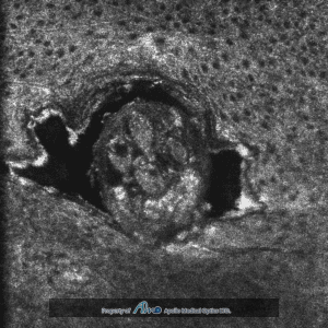
High resolution imaging
Cellular imaging has also been carried out using a confocal microscopy technique with a high numerical aperture (NA) objective. Another reason to use a high NA objective is to have an acceptable axial resolution (i.e. five to ten microns); however, this approach limits the imaging depth of an optical imaging system. Full-field OCT can achieve high axial resolution with a lower numerical aperture compared to confocal microscopy because axial sectioning is performed with both confocal gating and coherence gating. ApolloVue® achieves one micron resolution in both axial and lateral directions and also provides both en-face and cross-sectional cellular images. (Fig. 3)
Fig. 3 Better resolution is achieved by combining confocal gating and coherence gating.
Crystal Fiber
Single-crystal fiber is used in high-power fiber lasers in place of normal glass fiber since it has higher thermal conductivity and reduced nonlinear effects. AMO’s ApolloVue® system utilizes the crystal fiber technique as a broadband light to achieve high resolution. The single-crystalline fiber cores, with an inner diameter of a few tens of microns, were grown in air with the LHPG method.
AMO use the crystal fiber in ApolloVue® ’s light source module, providing improved OCT performance, including:
Continuous wave output: minimum invasiveness
High brightness: high lateral resolution, high lateral image quality and high imaging speed
Broadband width: one micron axial resolution
Gaussian spectrum: good axial image quality
Need help?
Please send your inquiries to our email address and we'll connect you to an expert.